


Now Reading: ECG 2D Echo TMT and Angiogram: Why Are These Heart Tests Necessary?
-
01
ECG 2D Echo TMT and Angiogram: Why Are These Heart Tests Necessary?
ECG 2D Echo TMT and Angiogram: Why Are These Heart Tests Necessary?

ECG
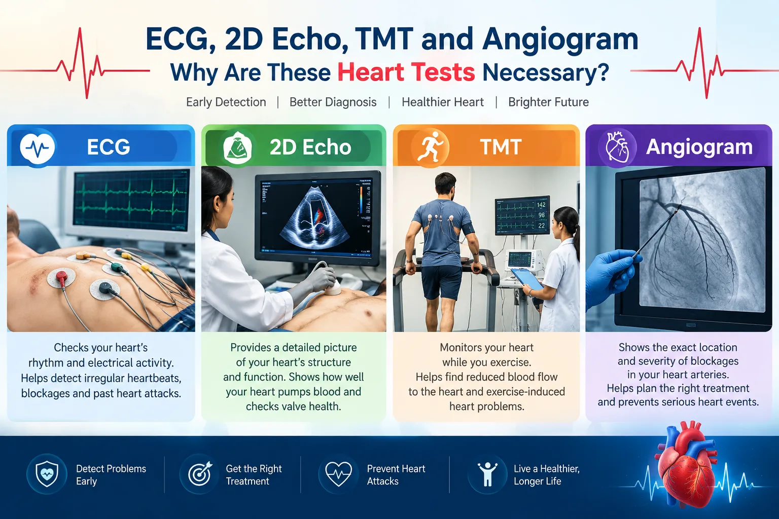
Doctors use different cardiac tests (ECG 2D Echo TMT and Angiogram) because no single test can assess every aspect of the heart. An ECG primarily evaluates the heart’s electrical activity, a 2D echocardiogram evaluates heart structure and function, a TMT evaluates the heart during exercise, and a coronary angiogram directly visualizes the coronary arteries.
These (ECG 2D Echo TMT and Angiogram) tests may be recommended when a person has symptoms such as chest discomfort, breathlessness, palpitations, dizziness or unexplained fatigue, or when there are risk factors for cardiovascular disease.
1. ECG Electrocardiogram (ECG, 2D Echo, TMT and Angiogram)
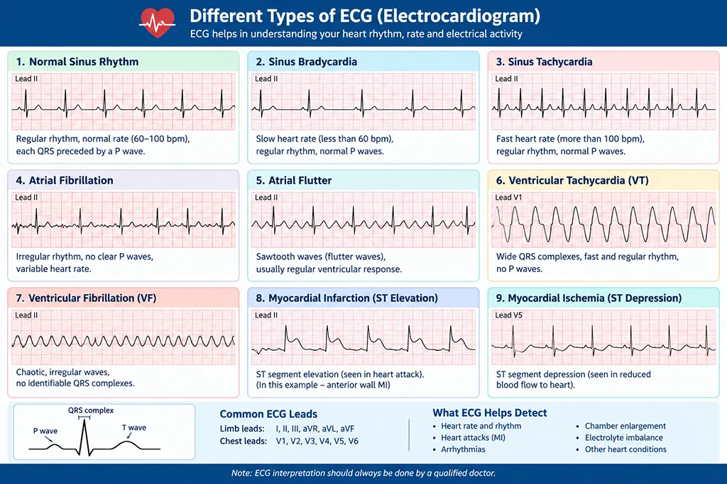
What is an ECG?
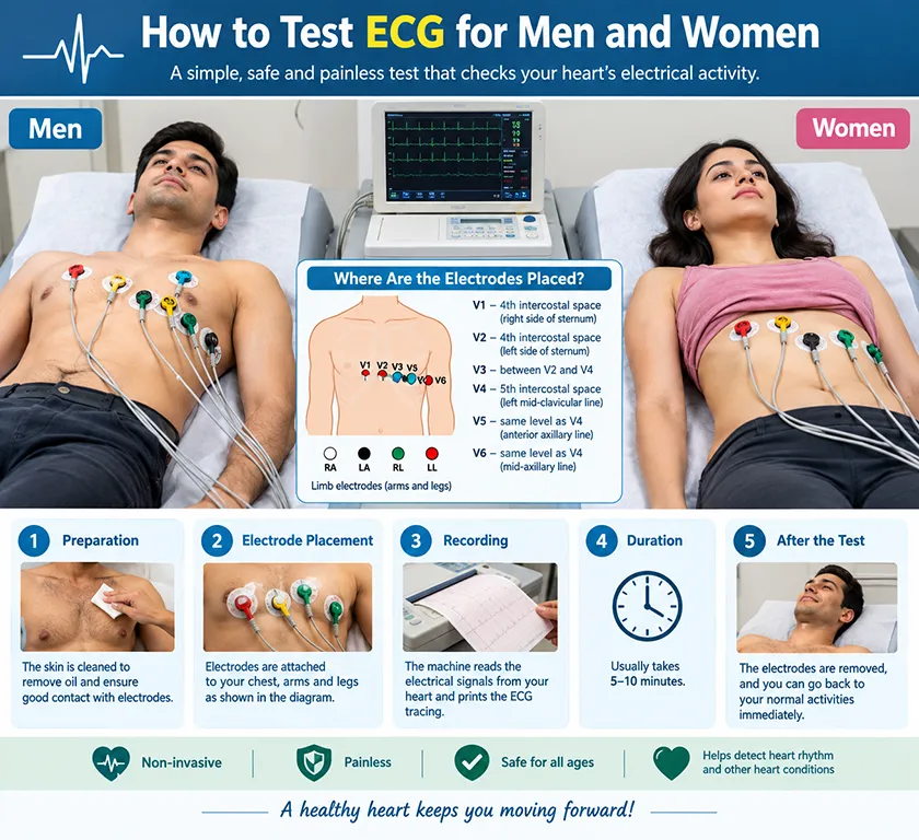
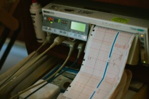
An electrocardiogram (ECG/EKG) is a quick, non-invasive test that records the heart’s electrical activity using electrodes placed on the chest and limbs.
What can an ECG detect?
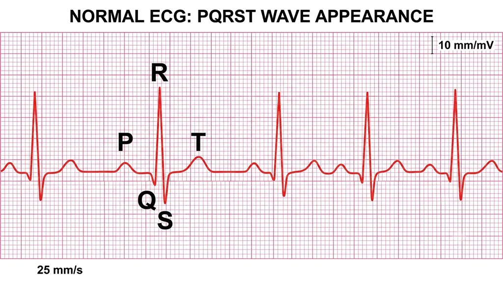
It can provide information about:
- Heart rate
- Heart rhythm
- Abnormal heart rhythms (arrhythmias)
- Evidence suggesting a previous heart attack
- Some patterns associated with reduced blood flow to the heart
- Conduction abnormalities
- Certain abnormalities associated with an enlarged or strained heart
Why is an ECG (ECG 2D Echo TMT and Angiogram)necessary?
An ECG is often one of the first cardiac tests performed because it is quick, inexpensive, painless and widely available.
A doctor may recommend an ECG for:
- Chest pain or pressure (ECG 2D Echo TMT and Angiogram)
- Palpitations (ECG 2D Echo TMT and Angiogram)
- Breathlessness (ECG 2D Echo TMT and Angiogram)
- Dizziness or fainting (ECG 2D Echo TMT and Angiogram)
- Unexplained fatigue (ECG 2D Echo TMT and Angiogram)
- Suspected heart attack (ECG 2D Echo TMT and Angiogram)
- Suspected arrhythmia (ECG 2D Echo TMT and Angiogram)
- Monitoring certain heart conditions or medications (ECG 2D Echo TMT and Angiogram)
Important limitation
A normal ECG does not rule out every heart problem. Some coronary artery problems or intermittent rhythm abnormalities may not appear on a resting ECG.
2. 2D Echo — Echocardiography

What is a 2D Echo?
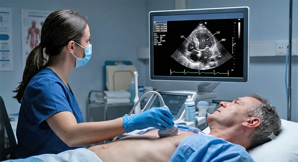
A 2D echocardiogram uses ultrasound waves to produce moving images of the heart.
Unlike an ECG, which primarily examines electrical activity, an echocardiogram examines the heart’s structure and mechanical function.
What does a 2D Echo evaluate?
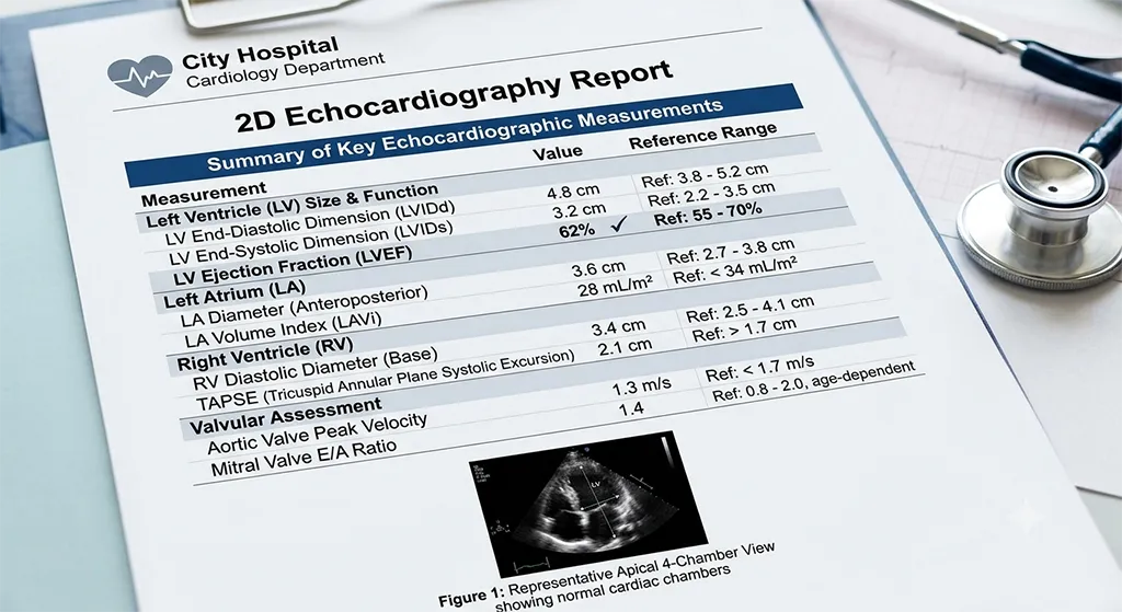
It can assess:
- Heart chambers (ECG 2D Echo TMT and Angiogram)
- Heart valves (ECG 2D Echo TMT and Angiogram)
- Heart muscle movement (ECG 2D Echo TMT and Angiogram)
- Pumping function (ECG 2D Echo TMT and Angiogram)
- Ejection fraction (EF) (ECG 2D Echo TMT and Angiogram)
- Heart-wall motion (ECG 2D Echo TMT and Angiogram)
- Blood flow patterns when Doppler is used (ECG 2D Echo TMT and Angiogram)
- Fluid around the heart (ECG 2D Echo TMT and Angiogram)
- Some structural abnormalities (ECG 2D Echo TMT and Angiogram)
Why is a 2D Echo necessary?
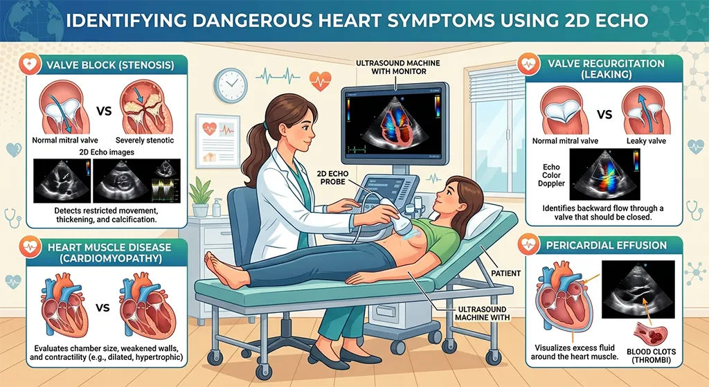
Doctors may recommend an echo when they need to determine whether the heart is:
Pumping properly → filling properly → structurally normal → functioning normally
It is particularly useful when evaluating:
- Heart failure (ECG 2D Echo TMT and Angiogram)
- Valve disease (ECG 2D Echo TMT and Angiogram)
- Cardiomyopathy (ECG 2D Echo TMT and Angiogram)
- Shortness of breath (ECG 2D Echo TMT and Angiogram)
- Abnormal ECG findings (ECG 2D Echo TMT and Angiogram)
- Certain congenital heart problems (ECG 2D Echo TMT and Angiogram)
- Previous heart attack (ECG 2D Echo TMT and Angiogram)
- Unexplained swelling or reduced exercise tolerance (ECG 2D Echo TMT and Angiogram)
What is ejection fraction?
Ejection fraction (EF) estimates how much blood the left ventricle pumps out with each contraction.
A reduced EF can indicate impaired pumping function, although heart failure can also occur when EF is preserved.
Therefore, EF should always be interpreted together with symptoms, examination and other test results.
3. TMT — Treadmill Test / Exercise Stress Test
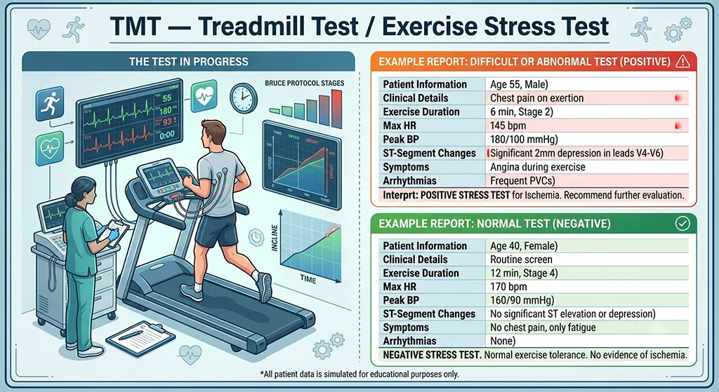
What is a TMT?
A TMT (treadmill test) is an exercise stress test.
The patient walks on a treadmill while healthcare professionals monitor:
- ECG
- Heart rate
- Blood pressure
- Symptoms
- Response to increasing exercise intensity
Why is a TMT necessary?
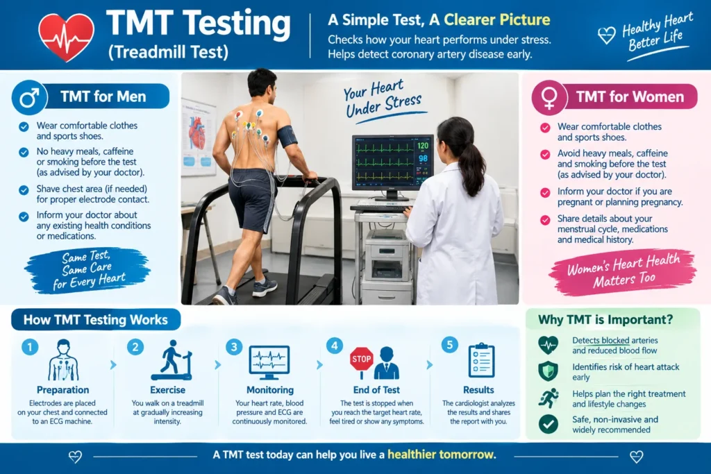
Some heart problems become apparent only when the heart has to work harder.
A TMT can help doctors assess whether exercise produces findings that suggest inadequate blood flow to the heart muscle, known as myocardial ischemia.
It can also provide information about:
- Exercise capacity
- Heart-rate response
- Blood-pressure response
- Exercise-induced arrhythmias
- Symptoms occurring during exertion
When might a doctor recommend it?
A TMT may be considered for selected patients with:
- Suspected coronary artery disease
- Exercise-related chest discomfort
- Breathlessness during exertion
- Unexplained reduced exercise capacity
- Certain abnormal ECG findings
- Assessment of exercise capacity in selected cardiac patients
Important limitation
A TMT is not suitable for everyone, and its accuracy depends on the patient’s circumstances. Doctors may choose other tests, such as stress imaging or CT coronary angiography, when those provide better information.
4. Angiogram Coronary Angiography
What is a coronary angiogram?
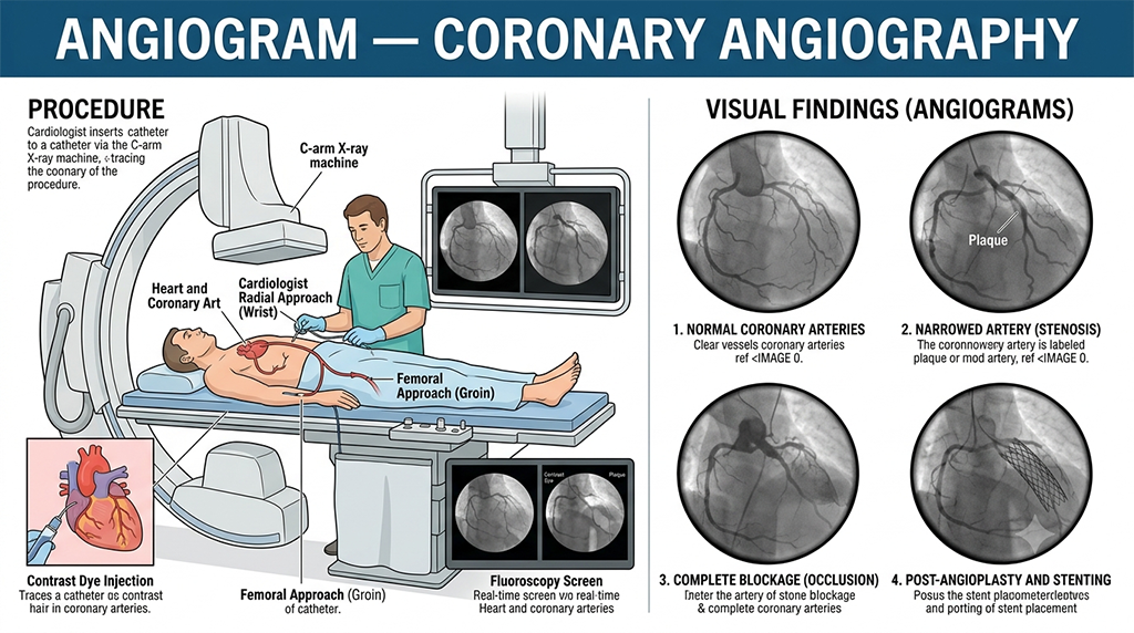
A coronary angiogram (coronary angiography) is an invasive imaging test used to visualize the coronary arteries.
A thin catheter is introduced through an artery, commonly through the wrist or groin, and contrast material is injected while X-ray images are taken.
What does an angiogram show?
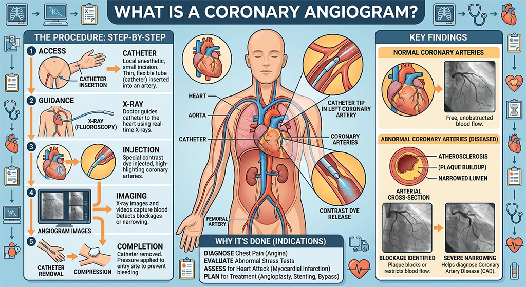
It can identify:
- Narrowed coronary arteries
- Significant blockages
- The location and severity of coronary artery disease
- The anatomy of coronary arteries
Why is an angiogram necessary?
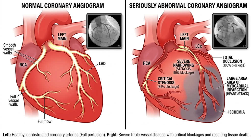
An angiogram may be recommended when doctors need detailed information about the coronary arteries, particularly when the likelihood of significant coronary artery disease is high or when other investigations indicate a potentially important blockage.
It may be used in situations such as:
- Suspected or confirmed acute coronary syndrome
- Significant symptoms suggesting coronary artery disease
- Abnormal results from other cardiac investigations
- Planning coronary angioplasty/stenting
- Planning coronary artery bypass surgery
Unlike a TMT, which assesses the heart’s response to exercise, coronary angiography provides direct images of the coronary arteries.
ECG vs 2D Echo vs TMT vs Angiogram
| Test | Mainly evaluates | Invasive? | Typical role |
|---|---|---|---|
| ECG | Electrical activity and rhythm | No | Initial cardiac assessment |
| 2D Echo | Structure and pumping function | No | Heart function, valves and structure |
| TMT | Response to exercise | No | Selected evaluation of exercise-related symptoms/ischemia |
| Angiogram | Coronary artery anatomy | Yes | Detailed assessment of coronary blockages |
A simple way to remember them
ECG → How is the heart’s electrical system working?
2D Echo → How is the heart structured and pumping?
TMT → How does the heart respond when it works harder?
Angiogram → Are the coronary arteries narrowed or blocked, and where?
Are all four tests necessary for everyone?
No.
A person does not automatically need an ECG, echo, TMT and angiogram simply because they have chest discomfort or are concerned about their heart.
The appropriate investigation depends on:
- Symptoms
- Age
- Medical history
- Blood pressure
- Diabetes
- Cholesterol
- Smoking history
- Family history
- Physical examination
- Initial ECG findings
- Previous heart disease
- Results of earlier tests
A cardiologist may start with an ECG and other basic assessments and then decide whether an echocardiogram, stress test, CT coronary angiography or invasive coronary angiography is appropriate.
When should you seek urgent medical attention?
Do not wait for a routine cardiac test if you develop new or severe chest pressure/pain, particularly if it is accompanied by:
- Shortness of breath
- Cold sweating
- Nausea or vomiting
- Fainting
- Severe weakness
- Pain spreading to the arm, shoulder, back, neck or jaw
These can be warning signs of a heart attack or another cardiovascular emergency. Seek emergency medical care immediately.
Key takeaway
These four tests answer different clinical questions. An ECG examines electrical activity, a 2D Echo evaluates structure and function, a TMT evaluates the heart under exercise stress, and an angiogram provides detailed visualization of the coronary arteries. The tests should therefore be selected based on the patient’s symptoms and clinical risk rather than performed routinely as a package.
Related Posts



Stay Informed With the Latest & Most Important News
Previous Post
Next Post










Advertisement

-
 01Boost your gut health and immune system naturally
 -
 02Regenerative Stem Cell Treatments in Orthopedics: A Complete Guide to Healing Joints, Bones, and Soft Tissues Naturally
 -
 03Can You Get Vitamin D3 During Sunrise or Sunset? A Scientific Guide
 -
 04Extraordinary Lasoda Glue Berry: Health Benefits, Nutrition, and Traditional Uses
 -
 05Country Eggs: Detailed Information, Nutrition, Benefits, Risks, and How They Compare With Farm Eggs
 -
 06Seasonal mushrooms and snake hole mushrooms: A complete guide
 -
 07The Ultimate Guide to Making Sesame Seed Milk: Recipe, Nutrition & Amazing Health Benefits